
The ultrasound shows an elarged pylorus (click image for arrows).
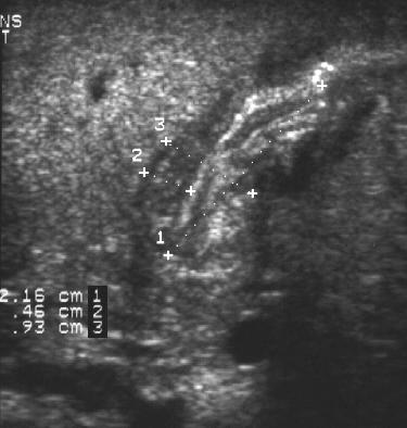
An ultrasound study is ordered to confirm the presence of pyloric stenosis.
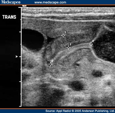
Hypertrophic pyloric stenosis. Long-axis view through the hypertrophied

2) Hypertrophic pyloric stenosis:

Pyloric stenosis in neonate:
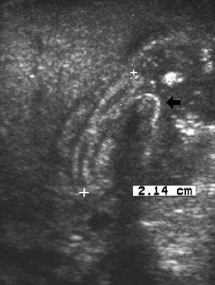
4 mm, and 14 mm, respectively is indicative of pyloric stenosis.

Pyloric stenosis
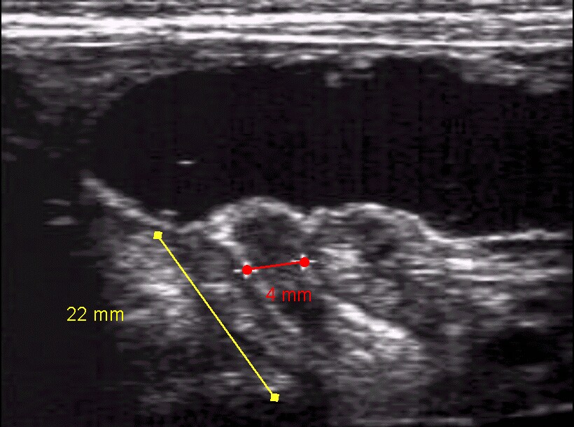
Longitudinal ultrasonogram of pyloric stenosis.

Pyloric stenosis - series : Normal anatomy

In pyloric stenosis, the muscles in this part of the stomach enlarge,
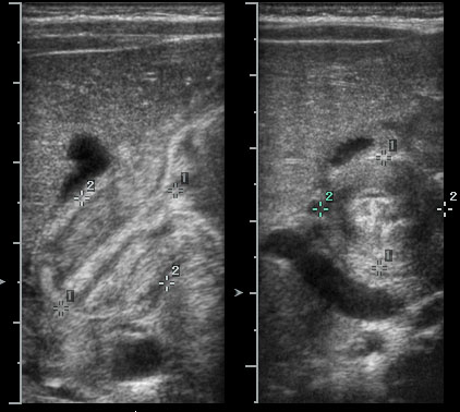
The pyloric muscle measures 5 mm thick and 21 mm

Pyloric ultrasound. <1> interval: length; <2> interval: muscle width

The validity of Ultrasound in Diagnosing Hypertrophic Pyloric Stenosis
Hypertrophic Pyloric Stenosis - MedPix™: 27207 - Medical Image Database and

Pylorus stenosis is usually apparent on ultrasound and responds to surgery.

Pyloric Stenosis flowchart. Pyloric Stenosis flowchart.gif

Pyloric stenosis is best diagnosed using ultrasound, which is ~99% sensitive
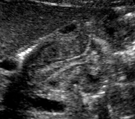
Longitudinal ultrasonogram in a patient with hypertrophic pyloric stenosis
((Pyloric Stenosis))
Ultrasound picture for the curious - 65K The outline shows the boundaries of
No comments:
Post a Comment